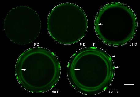
Fig. 4.
Long-term GFP labeling reveals the lamellar substructure of the lens. Lenses from Cre-ER™;Z/EG mice were examined at intervals (6-170 days) after tamoxifen injection. Optical sections were obtained through the lens equator. In this orientation, fiber cells are seen in cross section. Six days after tamoxifen treatment (6 D), scattered GFP-expressing fiber cells are observed near the lens surface. By 16 days (16 D), the fiber cells have become internalized and the innermost GFP-expressing cells are connected by the LMDP. Consequently, GFP is beginning to diffuse from the cytoplasm of expressing cells into the cytoplasm of non-expressing neighbors. The spread of fluorescence is largely between cells located in the same lens stratum, resulting in the formation of a fluorescent annulus which becomes better defined at later ages (arrowed in 21 D, 80 D, 170 D). At later ages (80 D and 170 D), secondary fluorescent rings (complete and incomplete) are present. These result from the periodic incorporation of clusters of highly fluorescent fiber cells (arrowheads in 170 D) into the lens syncytium. Scale bar: 500 μm.

