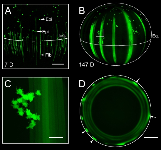
Fig. 5.
Age-dependent changes in clonal expansion of fiber cell progenitors accounts for the presence of secondary rings. (A) Maximum intensity projection of the lens equatorial region from a 29-day-old Cre-ER™;Z/EG mouse 7 days (7 D) after tamoxifen treatment. The position of the lens equator (Eq.) and individual GFP-expressing epithelial cells (Epi) is indicated. Note that GFP-positive epithelial cells are apparently randomly distributed in the lens epithelium and many, especially those located near the equator, are present as cell pairs. GFP-expressing fiber cells (Fib) are evenly distributed in the cortical lens tissue beneath the equator. (B) Maximum intensity projection of the lateral aspect of a lens from a Cre-ER™;Z/EG mouse 147 days (147 D) after tamoxifen treatment. Immediately anterior to the lens equator, clusters of GFP-positive epithelial cells are present (one cluster is boxed in B and shown at higher magnification in C). Diffusely labeled bundles of fiber cells are present interspersed with unlabeled fibers, resulting in a characteristic striped fluorescence pattern. (C) Clusters of GFP-positive epithelial cells contain 20-30 cells. (D) Equatorial optical sections of the lens at 147 days reveal the spatial relationship between the fluorescent fiber bundles and the pattern of secondary fluorescent rings. Near the periphery, discretely labeled bundles of GFP-positive cells (typically containing 20-30 fluorescent fiber cells) are present (arrowheads). As the fiber bundles are overlaid with more recently differentiated cells, GFP begins to diffuse into the cytoplasm of neighboring cells located in the same stratum of the lens as the GFP bundles (arrows). Diffusion of GFP throughout an entire stratum results in the formation of secondary fluorescent rings. Scale bars: 250 μm (A); 500 μm (B,D); 50 μm (C).

