Fig. 8.
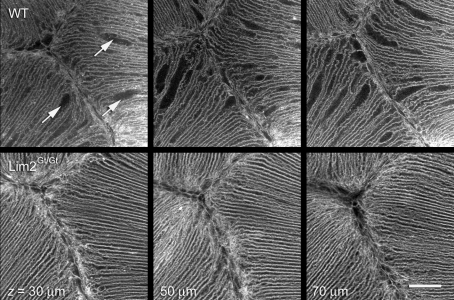
Conduits for intercellular diffusion of macromolecules might be Lim2-dependent cell fusions. Living, intact lenses were imaged by confocal microscopy following incubation in the lipophilic fluorescent dye, FM4-64. The ordered arrangement of the fiber cells in the vicinity of the anterior suture is evident but, in wild-type lenses, numerous cellular dilations are also present (arrows). Dilations are distributed throughout the tissue volume in this region, as shown by three representative sections from a z-stack collected beneath the anterior pole of the lens. A similar region (note the presence of the Y-shaped suture in each case) from a Lim2Gt/Gt mouse lens is shown in the lower panels. Cellular dilations are not present in the Lim2Gt/Gt lens. Scale bar: 50 μm.

