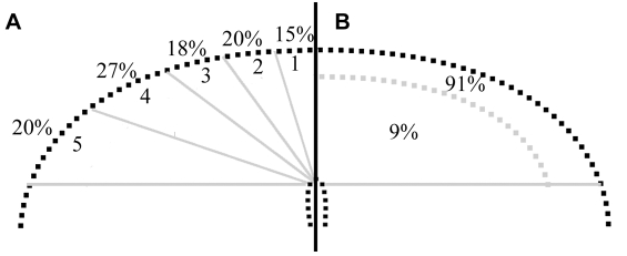
Fig. 7.
The surface position and apical-basolateral location of neural tube cell nuclei prior to an EMT. (A) Diagram of the dorsal neural tube showing the percentage of neural crest cells that emigrated from a given region of the neural tube. (B) The percentage of neural tube cell nuclei within a given apical-basal domain 1 hour prior to cell emigration.

