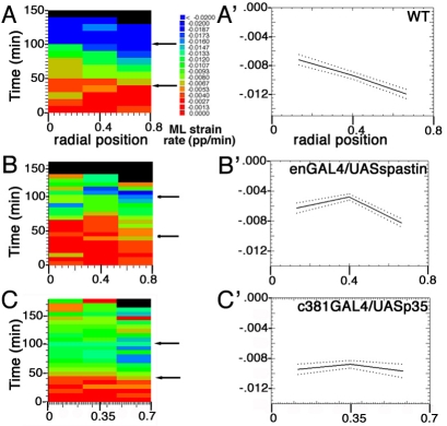
Fig. 5.
A gradient of AS cell contraction from the leading edge towards the dorsal midline depends on the zippering of the epidermis. (A-C) Proportional rates of change of cell shape in the ML orientation summarized by radial location. Cells at both canthi are removed from this analysis because they are known to be directly affected by zippering. Data are pooled from four wild-type (A), four enGal4/UAS-spastin-EGFP (B) and three ASGal4/UASp35 (C) Drosophila embryos. Note the `stair-like' distribution of shape strains in A, indicating that external cells are contracting their apical surface areas faster and earlier than central cells. (A′-C′) The same data as in A-C presented as time averages for 40-100 minutes after the onset of DC (arrows in A-C). In enGAL4/UAS-spastin embryos, there is only a peripheral gradient of apical contraction, whereas in ASGal4/UAS-p35 embryos there is no gradient of apical contraction along the radial axis (all cells contract at approximately the same rate).

