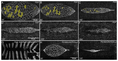
Fig. 6.
Zippering defects in mys mutant Drosophila embryos is not a consequence of defective filopodia, lack of cell extrusion or attachment to the yolk. (A-F) Still images from a time-lapse movie of a mys mutant embryo carrying the ubiECadGFP transgene (see Movie 4 in the supplementary material) at 20, 80, 140, 190, 220 and 250 minutes after the onset of DC. After the epidermis and the AS tear apart, the AS keeps on contracting (D-F). (G) Image from a time-lapse movie of a mys mutant embryo carrying the enGal4 and UASactinGFP transgenes (see Movie 5 in the supplementary material). Dorsal-most epidermal cells form filopodia at their dorsal end (inset). (H,I) Still images from a time-lapse movie of a P0180/UAS-TorsoβPScyt embryo carrying the ubiECadGFP transgene (see Movie 6 in the supplementary material) at 80 and 140 minutes after the onset of DC. When expressed in wing imaginal discs, this construct produces blistering of the wing (Dominguez-Gimenez et al., 2007) (data not shown).

