Fig. 5.
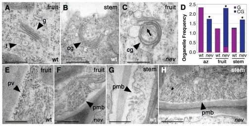
The fruit and stems of nev flowers also show membrane trafficking defects. (A-C,E-H) Transmission electron micrographs of cells in the fruit wall and pedicel (stem) of wild-type and nev flowers (stage 16). (A-C) Circularized multilamellar structures are enriched in nev fruit and stem cells (C,D) by comparison with the linear Golgi cisternae of wild-type cells (A,D). Rarely, Golgi cisternae with a curved appearance were observed in wild-type cells (B,D). Vesicular structures resembling the TGN of wild-type cells (A) are often observed in the vicinity or inside the multilamellar structures of nev cells (C, arrow) (73%, n=15). (D) Frequency of Golgi with a wild-type appearance (G, purple) and circularized multilamellar structures (CG, blue) per cell in sections of nev and wild-type sepal AZ regions, fruit walls and pedicels. For each type of tissue analyzed, n (cells) ≥15. Statistical differences between nev and wild-type tissues are indicated by asterisks (Fisher's exact test, P<0.0001). (E-H) Large paramural bodies are found in nev fruit (F) and stem (H) cells. Paramural vesicles and paramural bodies were also observed in wild-type fruit (E) and stem (G) cells. cg, circularized multilamellar structures; g, Golgi cisternae; pmb, paramural body; pv, paramural vesicles; t, TGN. Scale bars: 0.5 μm.

