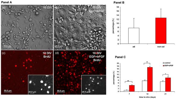
Figure 3.

Proliferative capacity of non-adherent HUCBmnf after 10 DIV with or without growth factors. Panel A: (a) brightfield photomicrograph of HUCBmnf cultured for 10 DIV without growth factors. The culture in (b) was treated with hEGF and hbFGF. BrdU+ cells were found in both the non-treated (c) and growth factor treated (d) cultures. The insets in (c) and (d) show BrdU labeled cells at higher magnification. Scale bar = 50 μm in (a)-(d), and 20 μm in inset of (c) and (d). Panel B: the proliferative capacity of the non-adherent cells tended to be greater than in the adherent HUCBmnf cells at 14 DIV (p = 0.06). Panel C: within the nonadherent fraction, treatment of the cultures with growth factors increased BrdU incorporation into the cells. Significant differences between the two groups were assessed by Student's t-test (*p < 0.05, **p < 0.001).
