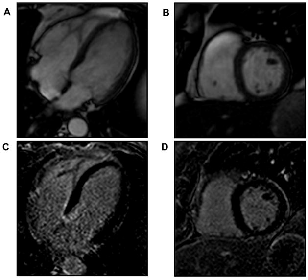
Figure 3.
The absence of late gadolinium enhancement (LGE) is evident in this patient with systemic amyloidosis but no cardiac involvement. Panels A and B illustrate steady state free precession images in the 4-chamber and short axis orientations respectively. Panels C and D are corresponding LGE images showing normal myocardial signal characteristics (nulling) without evidence of LGE.

