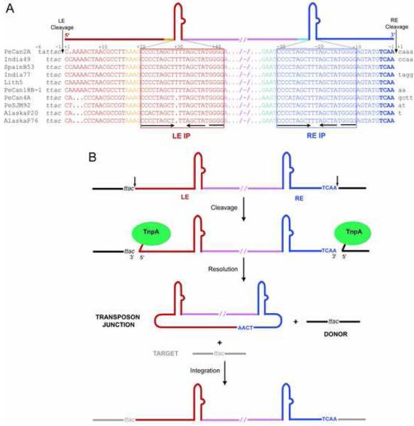
Figure 1. IS608 Transposition.
(A) Alignment (adapted from Kersulyte et al. [2002]) of the termini and flanking sequences of IS608 elements from Helicobacter pylori. The strain is indicated on the left; all the work reported here was done with IS608 from PeCan2A (top line). Flanking DNA is in black, bases of the left end (LE) are shown in red and orange, and bases of the right end (RE) are shown in shades of blue. Boxed sequences underlined with inverted arrows delineate the Imperfect Palindromes (IP) at each end. The bases are numbered such that, at each end, the cleavage site is between base−1 and base+1 where bases 5′ of the cleavage site are negative, and those 3′ are positive. (B) Model of single-stranded transposition from Guynet et al. (in press). Intermediates in the reaction are a circular transposon junction and a precisely sealed donor backbone.

