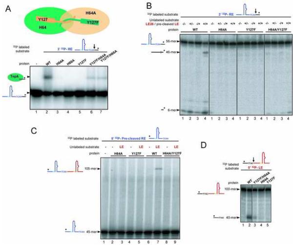
Figure 6. Mixed Mutant Dimers Suggest Transposon End Cleavage is in Trans and Resolution is in Cis.
(A) Results of RE cleavage assays using mixed active site point mutants. The radiolabeled oligonucleotide is detected. (B) RE cleavage and donor backbone formation by mixed active site mutants was assessed by mixing a RE cleavage substrate (56-mer) with a LE flank (40-mer). The product of RE cleavage is a 6-mer, and the 46-mer is the sealed donor backbone that forms between the LE flank and the RE cleavage product. (C) Transposon junction formation by mixed active site mutants was assessed by mixing a LE cleavage substrate (100-mer which is cleaved to yield a 60-mer transposon LE) with a precleaved transposon RE (45-mer). The transposon junction is formed by the WT protein (lane 7) but not by mixed active site mutants (lane 9). (D) LE cleavage is catalyzed by mixed active site mutants (lane 3), but not by the single point mutants (lanes 4 and 5).

