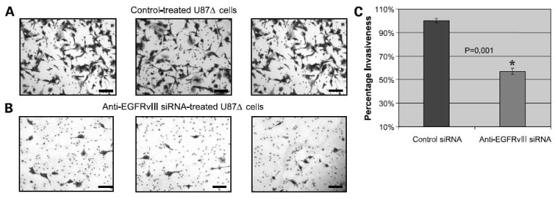
Figure 4. Anti-EGFRvIII siRNA reduces the invasiveness of glioma cells.

A, light microscopy (×40) images of the density of invaded U87Δ cells on the third day following control electroporation. Invasiveness was evaluated by a Matrigel-based cell invasion assay. Representative of three independent experiments done at 72 h post-electroporation. Cells were fixed and stained with crystal violet. Bar, 300 μm. B, light microscopy (×40) images of the density of invaded U87Δ cells on the third day following electroporation with anti-EGFRvIII siRNA. Invasiveness was evaluated by a Matrigel-based cell invasion assay. Bar, 300 μm. C, quantification of invaded U87Δ cells following electroporation with and without anti-EGFRvIII siRNA. This was achieved by dissolving U87Δ cells in NP-40 solution and performing spectrophotometry.
