
Fig. 1.
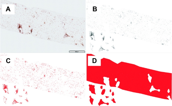
Immunohistochemical analysis using ImageJ software. (A) Representative image obtained via CD3 immunohistochemistry using the Olympus Dotslide system. The scale bar (500 μm) allows absolute areas to be calculated. (B-D) Analysis process for membranous staining. (B) ImageJ-enhanced 8-bit black and white image with portal tracts cut out to allow separate analysis of both lobular and portal regions. (C) Positive immunohistochemistry defined in red using a primary antibody-dependent standardized threshold, the area of which provides the numerator for positive immunohistochemical staining. (D) Threshold that gives a total area for both portal tract and lobular regions; the denominator. Analysis of nuclear staining is identical to membranous immunohistochemical staining, except that the immunohistochemical numerator is the number of positive cells. After a watershed is applied to separate overlapping cells, ImageJ calculates the number of positively stained cells using operator-determined shape and size characteristics.