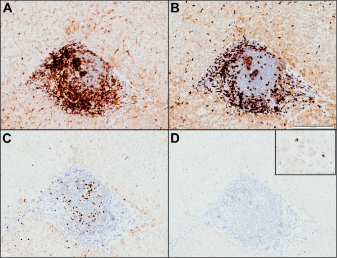
Fig. 3.

Immunohistochemistry of the portal inflammatory infiltrate from a patient with anti–HCV antibody but negative for HCV RNA. Formalin-fixed, paraffin-embedded tissue was stained for (A) CD4, (B) CD8, (C) Mcm-2, and (D) perforin. Scale bars: (B) 200 μm; (D) 50 μm (inset). Portal tracts are rich in CD3-positive cells (not shown), which are more often CD4-positive than CD8-positive. These cells express Mcm-2 and perforin rarely.
