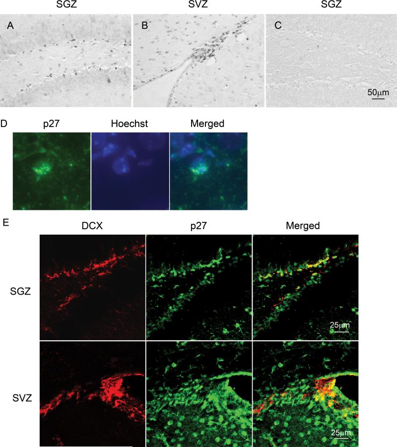
Figure 1. Expression of p27 protein in normal dentate gyrus and ventricular areas.
The histological sections of brain tissues derived from 8−10 weeks old normal mice (C57BL/6). The paraffin sections were stained with anti-p27 antibody and analyzed by microscopy. The representative images show that p27 positive cells reside at SGZ of dentate gyrus (A) as well as SVZ (B). A negative control for this staining (without primary antibody) was used and there was no staining on it (C). The scale bar is 50μm. D: High magnification of p27 and 4'-6-Diamidino-2-phenylindole (DAPI) staining in SGZ shows p27 staining is colocalized with nucleus. E: The normal brain sections were stained with anti-p27 and anti-doublecortin antibodies and analyzed by confocal microscopy. Upper and lower panels show the staining patterns in SGZ and SVZ respectively.

