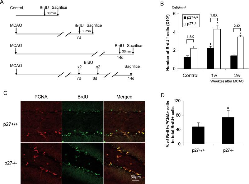
Figure 5. Increased proliferation of neural progenitors in p27−/− mice after ischemia.
The mice were subjected to 20-min MCAO, and BrdU was injected at either day 7 or day 14 (single dose). Mice were sacrificed 30 min thereafter. Control mice were sham-operated mice and receiving the same dose of BrdU as the ischemic mice. A. Schema of experimental design. B: The BrdU+ cells in SGZ were counted in 5 sections and calculated as described in Methods. *: p<0.05. C: The mice were subjected to 20-min MCAO, and BrdU was injected at day 7and 8. Animals were sacrificed at day 14 after MCAO. The coronal sections were stained with anti-BrdU and PCAN antibodies. The pictures show PCNA (red), BrdU (green) and merged staining in p27+/+ mice (upper penal) and p27−/− mice (lower penal). D. Quantitative data of Fig. 5C. BrdU+ cells and BrdU+/PCNA+ cells were counted. A higher percentage of BrdU+/PCNA+ cells in total BrdU+ cells were found in p27−/− mice compared with their littermates. The data is shown as mean ± SD, *: p<0.05.

