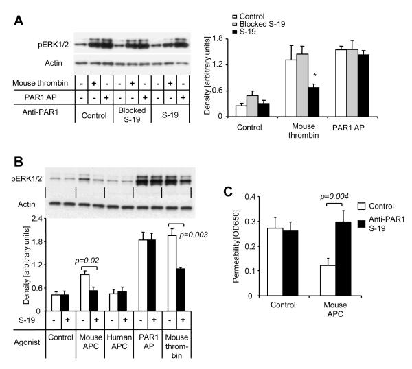
Figure 3. Anti-PAR1 S-19 blocks PAR1 signaling and APC-mediated barrier protection in mouse endothelial cells.
A) ERK1/2 phosphorylation was analyzed after 7 min incubation with mouse thrombin (1 nM) or the PAR1 agonist peptide TFLLRNPNDK (PAR1 AP; 20 μM). Where indicated anti PAR1 S-19 (25 μg/ml) alone or blocked by preincubation with excess of immunizing peptide was added 15 min prior to the agonists. A representative blot is shown on the left side, quantitative analyzes of S-19 immunoreactive bands are given in the right part of the figure (means±SEM) and ANOVA reviled that S-19 only significantly affects the thrombin response (*P<0.05). B) ERK1/2 phosphorylation in response to the indicated agonists (7 min) was analyzed in the absence or presence of S-19. APC was used at 20 nM. Representative blot shown in the upper part, quantification (means±SEM) of 4 independent experiments in the lower part, P value indicated. C) Subconfluent murine endothelial cells (b.End3) were incubated for 3 h with mouse APC (20 nM) in the absence or presence of S-19 in a dual chamber system followed by analysis of permeability. Means±SEM, n=9; P value indicated.

