Figure 4.
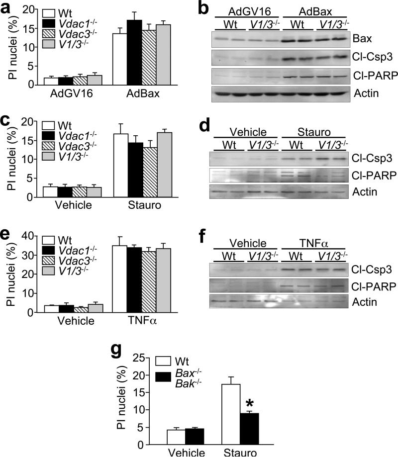
Cell death in Vdac-deficient MEFs. (a) Propidium iodide (PI) staining in wildtype, Vdac1-, Vdac3-, and Vdac1/Vdac3-null MEFs infected with adenoviruses encoding inducible Bax (AdBax). (b) Western blotting for Bax, cleaved caspase-3 (Cl-Csp3), cleaved PARP (Cl-PARP), and actin was performed in parallel. (c) PI staining in wildtype, Vdac1-, Vdac3-, and Vdac1/Vdac3-null MEFs treated with 300 nM staurosporine (Stauro) for 18 h. (d) Western blotting for cleaved caspase-3, cleaved PARP, and actin was also analyzed in parallel. (e) PI staining in wildtype, Vdac1-, Vdac3-, and Vdac1/Vdac3-null MEFs treated with 3 ng/mL TNFα plus 0.1 μg/mL actinomycin-D for 18 h, (f) and corresponding western blotting for caspase-3 and PARP cleavage. (g) PI staining in wildtype and Bax/Bak double-null MEFs following 300 nM staurosporine (Stauro) treatment for 18 h. All graphs show the average of 4 independent experiments. Error bars are s.e.m., and the asterisk denotes P<0.05 versus wildtype.

