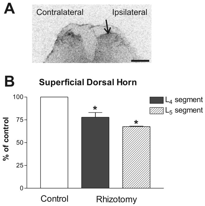
Fig. 3.
Representative autoradiogram of the AT1 receptor binding in the superficial dorsal horns of the lower lumbar spinal cord segment after unilateral selective dorsal rhizotomy. (A) Autoradiogram showing the AT1 receptor binding on the ipsilateral (lesion) and contralateral (intact) side. Arrow points to the superficial dorsal horn on the lesioned side. (B) Quantitative autoradiography of the AT1 receptor binding. Values are means ± SEM from groups of four animals, measured individually as described under Materials and Methods, and expressed as percentage of the control. *p<0.05 vs. contralateral side (control). Scale bar = 1 mm.

