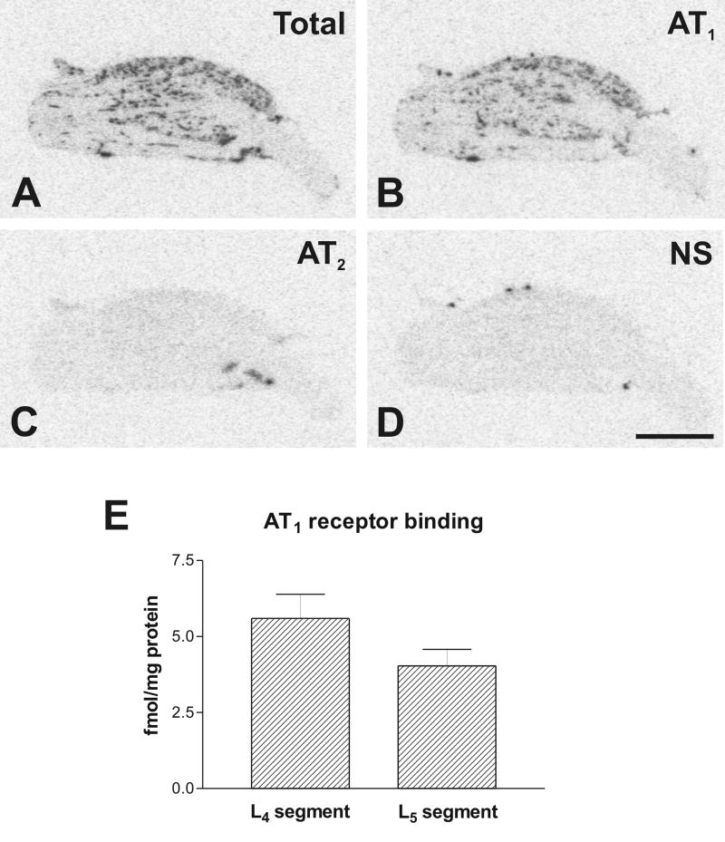
Fig. 4.
Representative pictures of Angiotensin II receptor binding in DRG from lower L4 lumbar level (A) Section showing total binding in the presence of [125I]-Sar1-Ang II. Consecutive sections showing binding as in A with addition of: (B) PD123319 to displace binding to AT2 receptors; (C) losartan to displace binding to AT1 receptors; (D) unlabeled Ang II displacing binding to AT1 and AT2 receptors. (E) Quantitative autoradiography of AT1 receptors in the DRGs. Values are means ± SEM from group of five animals, measured individually as described under Materials and Methods, and expressed as fmol/mg protein. Scale bar is 1 mm. NS, non specific.

