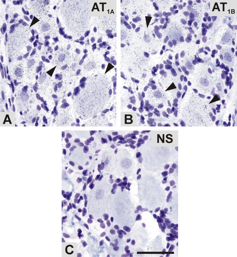
Fig. 6.
Bright-field microphotographs representing emulsion autoradiography of AT1A and AT1B mRNA in the DRG, as revealed by in situ hybridization. Aggregations of silver grains of antisense AT1A (A) and AT1B (B) riboprobes were detectable in the DRG neuronal cells (arrowheads). (D) AT1A sense control indicating the level of non-specific hybridization. Scale bar= 50μm. NS, non specific.

