
Fig. 3.
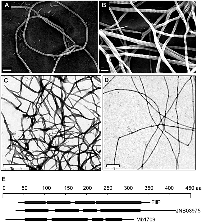
In vitro filaments formed by rod-domain proteins of actinomycetes. A and B. Scanning electron micrographs of filaments formed by S. coelicolor protein SCO5396 (FilP) in 50 mM TrisHCl at pH 7.0. A smooth non-branching filament is shown in A and the striated branching filaments are shown in B. C and D. Transmission electron micrographs of negatively stained filaments formed by Janibacter sp. protein JNB03975 and by M. bovis protein Mb 1709 respectively. E. Schematic representation of the various rod-domain structures of the proteins in A-C. Size bars represent 200 nm for A and B, and 1 μm for C and D.