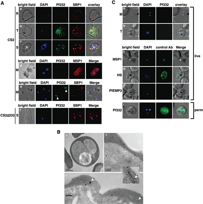
Fig. 6.

Detection of Pf332 in P. falciparum parasites throughout the blood-stage life cycle.
A. Localization of Pf332 in ring stage-infected erythrocytes (R) (a), trophozoite-infected erythrocytes (T) (b) and schizont-infected erythrocytes (S) (c). Each panel contains from left to right: bright field, DAPI-stained nuclei, anti-Pf322, anti-SBP1 and overlay of all four. Free merozoites (M) are shown in d. d is as above except that the fourth panel was labelled with anti-MSP1 antibodies. Panel row e contains free merozoites and material positive for both Pf332 and SBP1 suggesting Maurer's clefts (white arrows) that were released during schizont rupture. Panel row f shows CS2ΔPf322 schizont-infected erythrocytes (S). The order of each panel is as described above.
B. Transmission electron microscopy performed on high pressure frozen and freeze substituted sections of CS2-infected erythrocytes. The sections were labelled with either rabbit or mouse anti-Pf332 antibody and show the distribution of Pf332 in the red blood cell cytosol (a and b) and on Maurer's clefts (c and inset).
C. Live IFA on CS2-infected erythrocytes to determine whether Pf332 is on the surface of merozoites and/or trophozoite-infected erythrocytes. The first column of each row shows a bright field image, the second column a nuclear stain (DAPI), the third column reactivity with mouse monoclonal anti-Pf332 antibody and the last column an overlay of the previous three columns. Panels a and b: reactivity of anti-Pf332 antibodies with free live merozoites (M) and trophozoite (T)-infected erythrocytes to test for surface localization. Panel c: control panel with free live merozoites labelled with anti-MSP1 antibodies. Panel d: control panel with live trophozoites labelled with human serum (HS) from an individual living in a malaria-endemic region. Panel e: control panel with anti-PfEMP3 antibodies with live trophozoite-infected erythrocytes. Panel f: IFA on trophozoite-infected erythrocytes permeabilized with Triton X-100 to prove reactivity of the mouse monoclonal anti-Pf332 antibody.
