
Fig. 5.
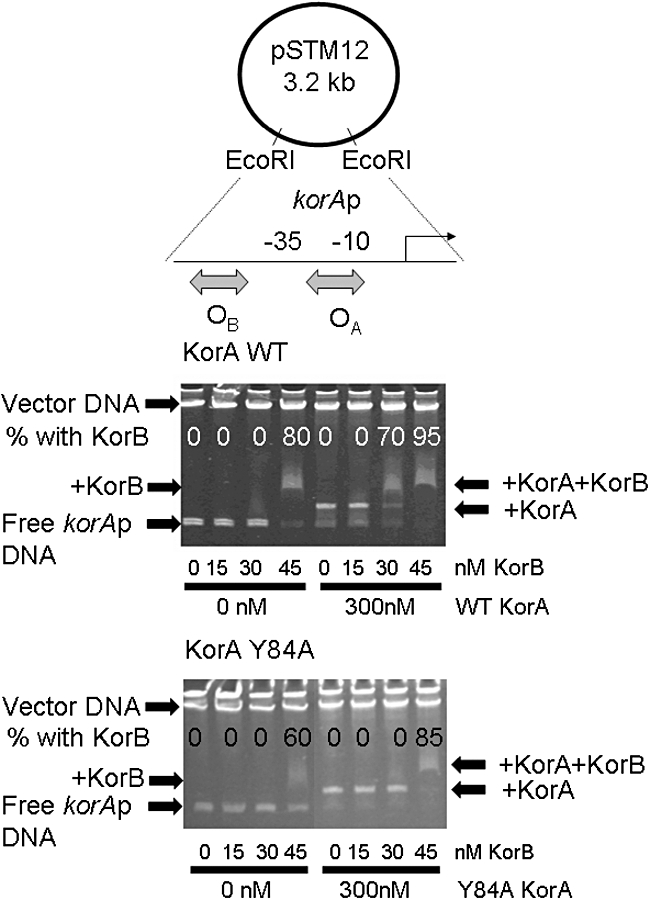
KorB electrophoretic mobility shift of a 220 bp korAp DNA fragment, in the absence and presence of KorA WT and Y84A. The korAp fragment was released from pSTM12 by EcoRI digestion – the 2.6 kb vector fragment runs at the top of the gel. The korA promoter includes KorA (OA: 5′-GTTTAGCTAAAC-3′) and KorB (OB: TTTAGCCGCTAAA-3′) sites separated by 20 bp. Each protein had previously been titrated separately to determine suitable concentrations. Proteins were added separately or together as described in Experimental procedures. The percentage of DNA fragment in the KorB–DNA complex, with or without KorA, is indicated on the gel. At 30 nM KorB the presence of 300 nM WT KorA results in > 80% retardation of the KorAp–KorA complex whereas the presence of 300 nM Y84A KorA has no effect.