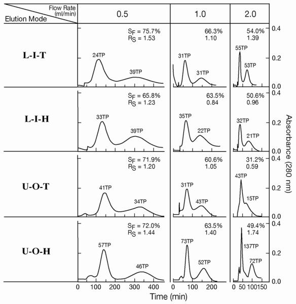
Fig. 5.
Protein separation by cross-pressed spiral tube assembly.
Experimental conditions are as described in Fig. 4. In L-I-T and L-I-H, the first peaks represent myoglobin and the second peaks, lysozyme while in U-O-T and U-O-H, the first peaks represent lysozyme and the second peaks, myoglobin.

