Fig. 3.
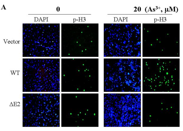
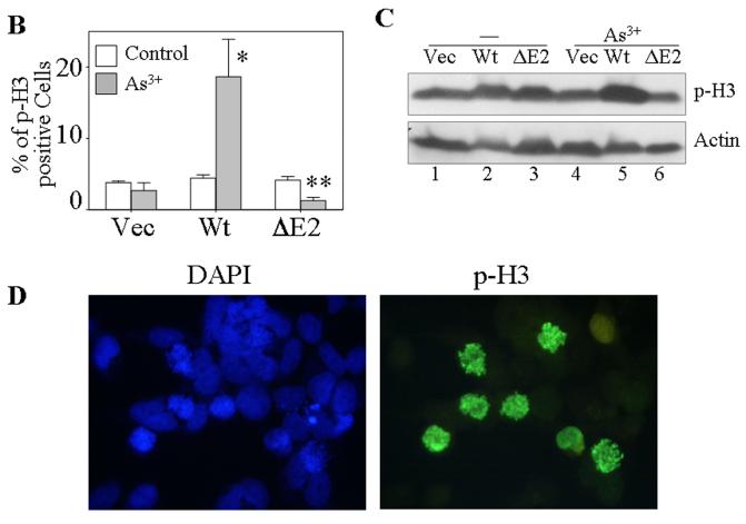
GADD45α1 is unable to sensitize As3+ induced mitotic arrest. A. The BEAS-2B cells stably expressing a control vector, GADD45α (WT) or GADD45α1 (ΔE2) were cultured in the absence or presence of 20 μM As3+ for 12 h. The cell mitosis was indicated by immunofluorescence staining using anti-phospho-histone H3 antibody (p-H3) and counter stained with DAPI to visualize the nuclei. B. Average percentage of the p-H3 positive cells of the vector-, GADD45α (wt)- or GADD45α1 (ΔE2)-transfected cells cultured in the absence or presence of 20 μM A3+ for 12 h. Data are means ± SD (n=3). *: p < 0.01; **:p < 0.005. C. Western-blotting analysis of the p-H3 in the cells transfected with the indicated vectors and cultured in the absence or presence of As3+. D. Morphological analysis suggested that As3+ treatment arrests cells in prometaphase.


