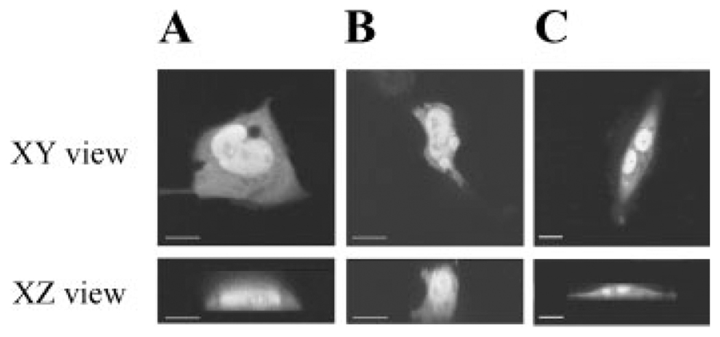
Fig. 2.
The distribution of GFP-VDR in proliferating BBe cells (A), differentiated BBe cells (B), and ROS 17/2.8 (AIG) cells (C). Confocal images of GFP-VDR distribution in a single optical section through the center of the cells in two axes: the XY plane and the XZ plane (reconstructed from multiple images in the XY plane).

