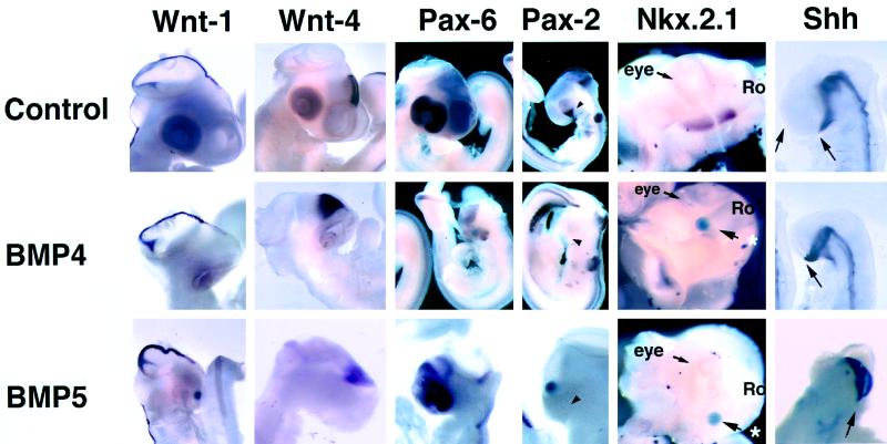
Figure 3.
In situ hybridization for dorsal and ventral markers in the forebrain of E4 embryos. Wnt-1 is expressed over the dorsal midline of the mesencephalon and diencephalon. Expression of Wnt-1 is normal or increased in embryos exposed to rBMP4 or rBMP5. Wnt-4 expression in the dorsal diencephalon expands ventrally in rBMP4-treated embryos compared with the control embryo. Pax-6, normally expressed in the dorsal telencephalon, is still expressed in the embryo exposed to rBMP4 and rBMP5, although the domain of expression is reduced in embryos exposed to rBMPs. In contrast to the dorsally expressed genes above, Pax-2 expression is completely absent in the ventral eye and ventral forebrain (arrowheads point to same region in each embryo). Nkx-2.1 expression normally seen in the basal forebrain is absent in embryos treated with rBMP4 and rBMP5. The embryonic head has been isolated and photographed from the ventral side. The eye (labeled) can be seen in each image and the rostral (Ro) end of the head is to the right in each image (∗ denotes the implanted bead). Shh expression in the ventral forebrain ends just rostral to the optic chiasm. The morphologically distinct basal telencephalon (arrows) is present rostral to the Shh expression domain. Embryos implanted with beads soaked in rBMP4 or rBMP5 show no morphologically identifiable basal telencephalon; the Shh expression domain comes up to the rostral limit of the ventral brain. All embryos were sectioned to confirm the whole-mount in situ hybridization staining patterns.

