Figure 6.
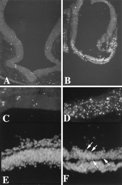
TUNEL assay for cell death. Relatively few cells were labeled by TUNEL in unoperated embryos or embryos implanted with a bead soaked in buffer (A, coronal section of forebrain, dorsal is up and ventral down; ×100). In contrast, a selective and extensive labeling of cells, indicating cell death, was found in the ventral telencephalon after exposure to rBMP5 or rBMP4 proteins (B, coronal section of E3 telencephalon, dorsal is up and ventral down, a bead soaked in rBMP4 protein was implanted at stage 11; ×100). At higher power, the difference between dorsal (C) and ventral (D) telencephalon is striking (both C and D from an embryo implanted at stage 9 with a bead soaked in rBMP5 protein and harvested on E4; ×400). 4′,6-Diamidino-2-phenylindole (DAPI) nuclear stain confirmed the TUNEL findings. (E) Field of dorsal neural tube of embryo exposed to rBMP4 protein shows all intact nuclei. (×400.) (F) A field from the ventral telencephalon of the same section as E shows numerous condensed nuclei (arrows) characteristic of cells undergoing apoptosis. (×400.)

