
Figure 4.
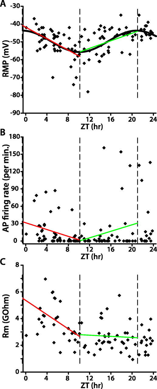
WT lLNv membrane excitability in LD is strongly rhythmic. A–C, Each data point in the scatter plots represents the RMP (A), spontaneous AP firing rate (B), or membrane resistance (C) of one lLNv at the indicated time of recording. In A, the sinusoidal curve represents the best fit of RMP as a function of time of day in LD (ZT, nonlinear regression; p < 0.0001). The three vertical dashed lines indicate the peak and trough of the fitted sinusoid of RMP versus ZT. Red and green lines depict the linear regression analysis of data within those two intervals (A, both red and green linear fits, p < 0.01; B, C, red linear fit, p < 0.05; green linear fit, p > 0.1).