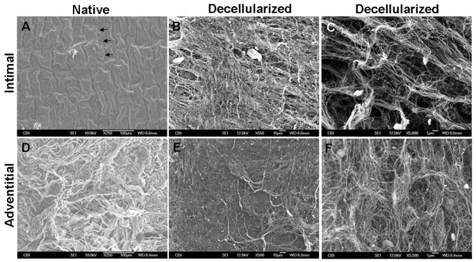
Fig. 3.
SEM of intimal and adventitial surfaces of native and decellularized arteries. (A) The intima of the native vessel shows a corrugated endothelial layer; a small tear shows exposed basement membrane underneath (arrows). (B) The intima of the decellularized artery shows exposed ECM. (C) Higher magnification reveals detailed pore structure. (D) The adventitial surface of the native artery has thick bundles of crimped collagen fibers. (E) The adventitia of the decellularized artery has a smoother surface, suggesting uncrimping of fibers. (F) Higher magnification of the decellularized adventitial surface shows detail of shallow pores and fibers.

