
Fig. 5.
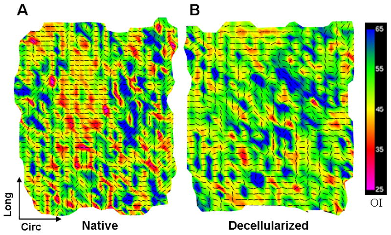
Fiber kinematics for non-pressurized arteries. Fiber architecture maps for (A) native and (B) decellularized arteries fixed at 0 mmHg. Black vectors represent the preferred local fiber orientations, and colors represent OI; warmer colors represent lower OI, which indicates a higher degree of alignment. Native arteries showed a high degree of fiber alignment towards the circumferential direction. Decellularized arteries had more random fiber orientation and lower alignment (higher prevalence of cooler OI colors), suggesting structural interruption.