
Fig. 1.
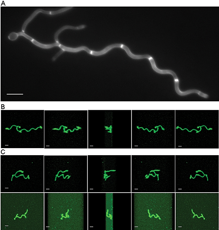
Induction of regular sinusoidal and helical hyphae of C. albicans by growth on surfaces in low-nutrient conditions. Sinusoidal curves were induced in hyphae of the control strain by growth on a poly-l-lysine-coated slide in 1% serum. Hyphae were stained with the chitin-specific brightener, Calcofluor White (A). Other hyphae were imaged using scanning confocal microscopy to view hyphal projections that had been stained with the lipophilic dye, FM4-64. Three-dimensional images were constructed and sequential frames are shown for hyphae rotated through the X-plane by 180° (B and C). When grown on 4% (w/v) agar containing 20% (v/v) serum, most hyphae grew as two-dimensional sinusoidal curves that could be shown to be growing in the plane of the substrate when observed as end-on projections (B). However, two examples are shown in C where hyphae formed three-dimensional helical loops that extended above the plane of the substrate. Scale bars = 10 μm.