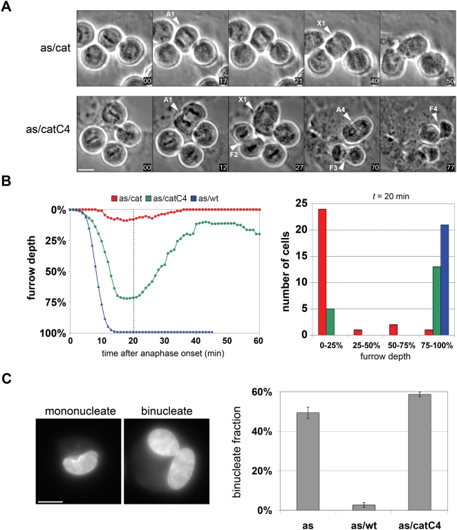
Figure 7. Tethering Plk1 to the centralspindlin complex rescues the initiation but not the completion of cytokinesis.
(A) Monastrol-synchronized cells were released for 60 min into drug-free medium, followed by addition of 3-MB-PP1 and initiation of timelapse imaging (time 00). Examples of cells that traversed anaphase (A) and either formed equatorial furrows (F) or failed to do so (X) are indicated (see also Videos S1 and S2). Scale bar, 10 µm. (B) Biphasic furrow dynamics in Plk1as/catC4 cells. At least 18 cells of each genotype were imaged as above, and the average depth of the furrow relative to the initial equatorial diameter at anaphase onset was calculated. Furrow trajectories in individual cells are provided in Figure S6. (C) Cytokinesis ultimately fails in Plk1as/catC4 cells. Cells were synchronized in prometaphase by monastrol block and release as above. Thirty minutes after monastrol release, 3-MB-PP1 was added to 10 µM for an additional 90 min to inhibit Plk1as activity during mitotic exit. Adherent cells were collected by trypsinization, fixed and stained with Hoechst 33352, and analyzed microscopically to determine the fraction of binucleated cells in each population (n = 3 determinations, 100 cells/sample; values are reported as means ± SEM). Scale bar, 10 µm.

