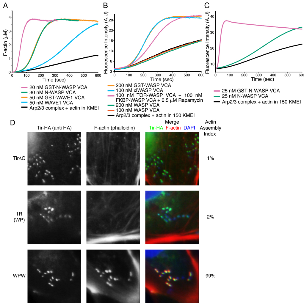
Figure 1. Dimerization Increases VCA Activity In Vitro and in Cells.
(A – C) Pyrene fluorescence measured during assembly of actin by Arp2/3 complex (black) plus the indicated components. Without rapamycin, 100 nM mTOR-WASP VCA + 100 nM FKBP-WASP VCA has activity comparable to 200 nM WASP VCA (not shown). (D) Clustered EspFu leads to actin rich pedestal formation, but requires the ability to recruit multiple WASP molecules. Cells expressing HA-tagged TirΔC, TirΔC-1R (WP) or TirΔC-WPW were treated with intimin-expressing E. coli, and stained with anti-HA antibody (green in merged image) and Alexa568-phalloidin (red in merged image). Colocalization is yellow in merged image. The fraction of transfected cells harboring at least five F-actin foci was quantified (Actin Assembly Index). Bacteria were visualized by DAPI staining (blue in merged image). Data represent mean from three independent samples of 30 cells each, standard deviation for all samples is 2%.

