Figure 3.
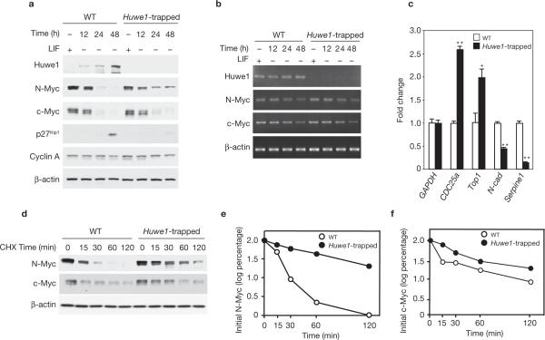
Genetic inactivation of Huwe1 impairs N-Myc degradation. (a) Wild-type and Huwe1-trapped ES cells were plated in the presence of LIF and 18 h later deprived of LIF for the indicated times. Lysates were analysed by immunoblotting using the indicated antibodies. (b) Parallel cultures were analysed for expression of Huwe1, N-myc, c-myc and β-actin by semi-quantitative RT–PCR. (c) Quantitative real-time PCR (qRT–PCR) analysis of the mRNA of selected Myc target genes in wild-type and Huwe1-trapped ES cells. Data represent mean ± s.e.m. (n = 3; *P < 0.01 **P < 0.001 Student's t-test). (d) Wild-type and Huwe1-trapped ES cells were treated with CHX for the indicated times 24 h after LIF deprivation. Lysates were analysed by western blotting using anti-N-Myc and anti-c-Myc antibodies. β-actin is shown as a control for loading. (e) Quantification of N-Myc from the experiment in d. (f) Quantification of c-Myc from the experiment in d. Full scans of immunoblots are shown in Supplementary Information, Fig. S9.

