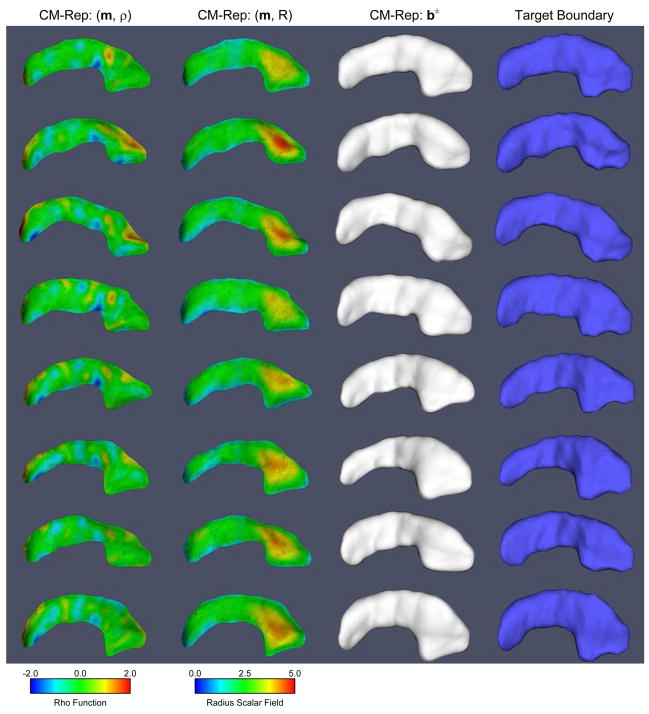
Fig. 5.
Examples of cm-rep models fitted to binary segmentaions in the hippocampus dataset. For each hippocampus, shown are the medial manifold colored by the ρ function (the right hand side of the biharmonic PDE), the medial manifold colored by the radius function R (the solution of the PDE), the boundary generated by inverse skeletonization (1), and the boundary of the segmentation to which the cm-rep was fitted.

