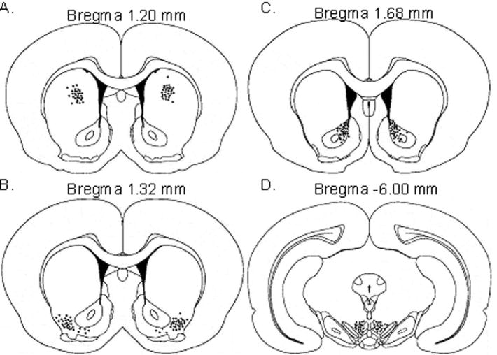
Figure 1. Cannula placements.
Injection sites for A) caudate putamen, B) lateral accumbens shell C) nucleus accumbens core, and D) ventral tegmental area. Locations of the tip of the cannulae are indicated by black circles. Figures reproduced from Paxinos and Watson (2007) with permission.

