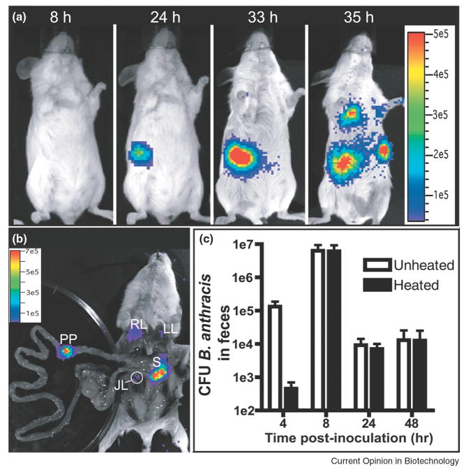
Figure 3.
Bioluminescence imaging to map germination of B. anthracis in gastrointestinal infection. B. anthracis spores (1×10⁸) were inoculated using a flexible plastic tube. (A) Infection of the lower digestive tract at the indicated times. Image series is representative of 23 mice. (B) Dissection of the mouse in (A) to confirm sources of luminescence. JL, jejunal lymph node; LL, left lung; PP, Peyer's patch; RL, right lung; S, spleen. (C) Total CFU of B. anthracis (unheated) or spores only (heated) were enumerated at the indicated times from the feces of mice inoculated intragastrically (mean ± standard error of the mean, n = 4). Vegetative bacteria were isolated from feces at four hours post-infection, indicating spore germination in the gastrointestinal tract. Adapted by permission from Public Library of Science: Glomski et.al., PLoS Pathogens, 3:0699-0708, copyright 2007.

