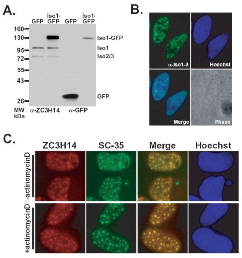
Figure 4. Endogenous nuclear ZC3H14 colocalizes with nuclear speckles.

A. Lysates from HeLa cells expressing either GFP or ZC3H14 isoform 1-GFP (Iso1-GFP) fusion protein were probed with polyclonal anti-ZC3H14 (α-ZC3H14) or anti-GFP (α-GFP) antibody. B. The localization of endogenous ZC3H14 (isoforms 1-3) in HeLa cells was determined by indirect immunofluorescence using the polyclonal anti-ZC3H14 antibody. Hoechst dye was used to stain DNA and indicate the position of the nucleus. The merge reveals that endogenous ZC3H14 (isoforms 1-3) is located in foci within the nucleus. A corresponding Phase image is also shown. C. ZC3H14 localizes to nuclear speckles in HeLa cells. ZC3H14 was detected with anti-ZC3H14 antibody (green) and nuclear speckles were labeled with anti-SC-35 antibody (red). Colocalization (yellow) is indicated in the merge panel. To assess whether ZC3H14 shows dynamics typical for nuclear speckle proteins, ZC3H14 was localized either prior to (-actinomycinD) or following (+actinomycinD) treatment with actinomycinD for 4 hr. As a control, the –actinomycinD sample was treated with DMSO, which is the solvent for actinomycinD. The merge image shows that nuclear ZC3H14 colocalizes (yellow) with nuclear speckles under all conditions examined.
