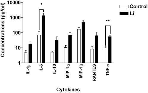
Figure 6. Leishmania infantum amastigotes promote secretion of IL-6 and TNF-α.
iDCs were either left unexposed (Control) or exposed to Leishmania infantum amastigotes (Li) for 24 h. Cell-free supernatants were collected and analyzed with a Bio-Plex assay. The results shown are representative of five separate experiments performed with different donors. Asterisks denote statistically significant differences from the uninfected cells (P<0.05; **, P<0.01).

