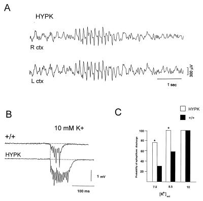
Figure 4.
HypK mice exhibit spontaneous electroencephalogram (EEG) discharges in vivo and have a lower threshold for epileptiform discharge in hippocampal slices. Cortical EEG traces were recorded from the right (R) and left (L) hemispheres in mice from the HypK high transgene copy number line (836) (12 adults 2–3 months of age) and HypK low transgene copy number line (12 age-matched adults). (A) Spontaneous bilateral spike and wave seizure activity was routinely observed. In 6 control FVB (+/+) mice (not shown), abnormal synchronous activity was never present. (B) Extracellular field recordings from the CA3 pyramidal cell layer reveal prolongation of K+-induced network discharge in HypK hippocampus. Upper trace is from a control FVB slice in 10 mM [K+]ext and the lower trace is from the HypK high transgene copy number strain, 836. Discharges from the 836 HypK mutant strain were significantly prolonged (≈20%; P > 0.05, t test). (C) Lower threshold for network bursting in HypK hippocampal circuits. Histogram indicating the number of slices, expressed as a proportion of the total, that showed spontaneous epileptiform activity at each [K+]ext. The proportion of slices exhibiting spontaneous activity in both 7.5 mM and 8.5 mM [K+]ext was approximately 2-fold greater in transgenic mice compared with control (P > 0.05, sign test).

