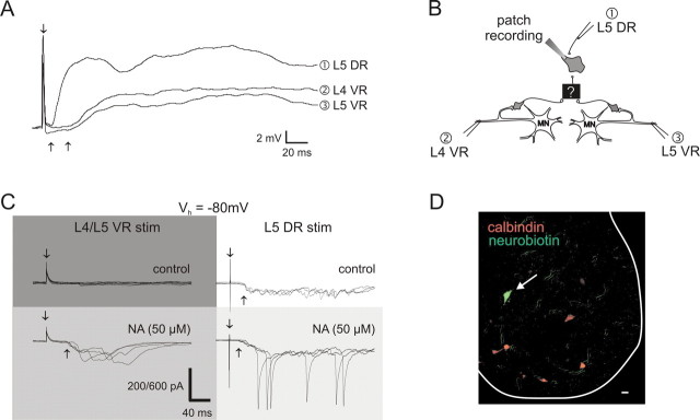
Figure 5.
Evidence for the existence of interneurons having actions expected from the properties of the evoked reflexes. A, Average of 10 whole-cell current-clamp recordings of a cell in the ventral horn near the motor pool that received short-latency excitation from dorsal root stimulation (500 μA, 500 μs) and long-latency excitation from ventral root stimulation (400 μA, 200 μs). B, Schematic diagram of the experimental setup illustrating the respective stimulating sites for responses (1–3) and hypothetical circuitry consistent with the observed actions. C, NA-dependent ventral-root-evoked synaptic actions in a spinal neuron. Membrane voltage was clamped at a holding potential of −80 mV (Vh of −80 mV). Under control conditions, the neuron received long-latency sensory input (300 μA, 200 μs) but no input from simultaneous L4/L5 ventral roots (top row; 300 μA, 200 μs). After addition of NA (50 μm), sensory input was facilitated and long-latency synaptic inputs after VR stimulation were unmasked (bottom row). The vertical scale bar represents 200 pA, except for the trial with dorsal root stimulation in the presence of NA in which it represents 600 pA. D, The neuron in C was Neurobiotin filled to identify its location (white arrow). Both the location and lack of calbindin immunostaining (a marker of Renshaw cells) demonstrate that the recorded neuron is not a Renshaw cell. Scale bar, 25 μm. DR, Dorsal root; VR, ventral root.

