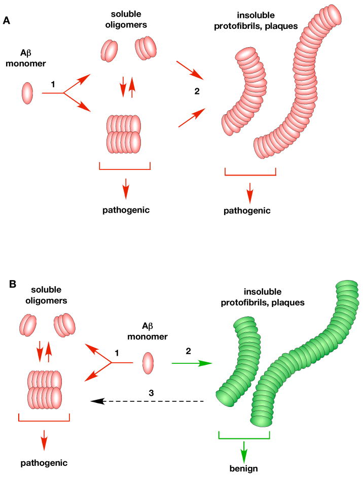
Figure 2. Potential pathways of Aβ oligomerization in vivo.
Almost nothing is known about how Aβ oligomerizes in vivo. One possibility (A) is that there is a single linear oligomerization pathway with Aβ monomers initially forming low molecular weight soluble aggregates (1). These oligomers further aggregate (step 2) into insoluble protofibrils, fibrils and plaques (2). In this view, both soluble oligomers and insoluble fibrils are considered to be pathogenic. An alternate possibility (B) is that there are two distinct pathways. A pathogenic pathway (1) that forms soluble oligomers (dimers/Aβ*56/ADDLs) which cause the disease, and a second non-pathogenic pathway (2) that leads to the formation of insoluble aggregates and plaques. The insoluble aggregates are thought to be benign, a view more consistent with the available data. It has also been suggested that the benign insoluble aggregates may slowly leach (3, dashed arrow) forming the pathogenic soluble oligomers (Meyer-Luehmann et al., 2008).

