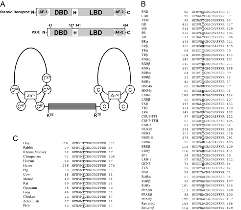
Fig. 1.
Thr57 in hPXR is conserved in other NRs. A, top panel showing the schematic comparison of the domain structures of a steroid receptor and hPXR. H, hinge region. Bottom panel shows the schematic representation of the two zinc finger motifs in the DBD of hPXR. The two zinc finger motifs are separated by a linker (from Lys62 to Arg76). The diagram was not drawn as per the scale in terms of number of residues. Note that Thr57 is located within the first zinc finger motif. B, Thr57 in hPXR is highly conserved among other human NRs. Of the 46 human NRs with DBD, 37 receptors (including all the classic steroid receptors) have a conserved T/S in the DBD. Nine receptors are exceptions, and they have an A instead of Thr/Ser at this position. Expansion of the abbreviated nomenclature for the receptors is as follows: CAR, constitutive androstane receptor; VDR, vitamin D receptor; GR, glucocorticoid receptor; MR, mineralocorticoid receptor; PR, progesterone receptor; AR, androgen receptor; ER, estrogen receptor; TR, thyroid hormone receptor; ROR, retinoid-related orphan receptor; LXR, liver X receptor; FXR, farnesoid X receptor; TR2, testicular receptor 2; TR4, testicular receptor 4; COUP-TF, chicken ovalbumin upstream promoter transcription factor; EAR-2, v-erbA-related; NURR1, NR-related 1; NOR1, neuron-derived orphan receptor 1; NGFI-B, nerve growth factor-induced clone B; ERR, estrogen receptor-related; SF-1, steroidogenic factor 1; LRH-1, liver receptor homolog-1; GCNF, germ cell nuclear factor; TLX, human homolog of the Drosophila tailless gene; PNR, photoreceptor cell-specific NR. C, Thr57 in hPXR is conserved in all the vertebrate species with a fully known PXR sequence. Chicken PXR is the only exception and has an S instead of T at that position.

