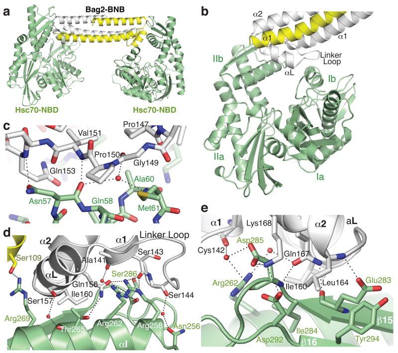
Figure 2.
Structure of the Hsc70-NBD:Bag2-BNB domain complex. (a) Asymmetric unit of the crystal, containing two Hsc70-NBD molecules and a Bag2-BNB dimer. (b) Closer view of the interface between the Bag2-BNB dimer and one of the Hsc70-NBD molecules. The BNB dimer contacts Hsc70-NBD subdomains Ib and IIb, largely through the Bag2 linker loop and αL helix. (c) Closeup of contacts between the Bag2-BNB linker loop and residues on subdomain Ib of the Hsc70-NBD. Dotted lines represent likely hydrogen bonds or salt bridges. (d) Closeup of contacts between the Bag2-BNB dimer and subdomain IIb of the Hsc70-NBD. (e) Closeup of additional contacts between Bag2-BNB and subdomain IIb of the Hsc70-NBD. This view is related to panel (d) by an approximate 180° rotation about the vertical axis.

