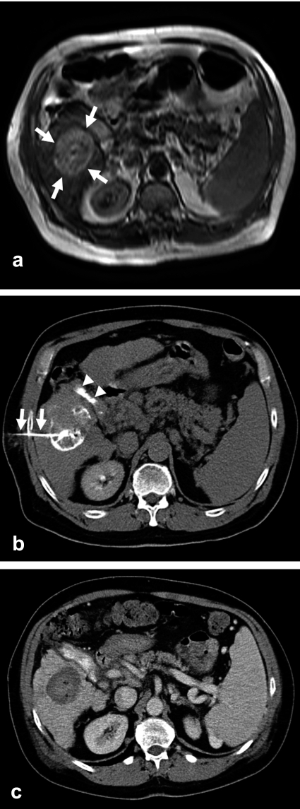
Figure 1.
The MRI scan of this 56-year-old man with longstanding hepatic cirrhosis due to hepatitis B and histologically confirmed hepatocellular carcinoma (HCC) reveals, in segments V/VI, a 4.6 cm sized tumor with nodular growth (a; arrows). The CT after tumor puncture with a dedicated 20 gauge needle with side holes (b; arrows) and injection of a mixture of ethanol and contrast medium shows a good distribution of contrast throughout the tumor. Gallstones are also seen (b;arrowheads). A CT obtained three months later shows the fully necrotic tumor as a parenchymal defect that does not take up contrast medium (c).

