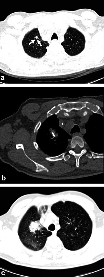
Figure 3.
CT images of a 72-year-old man with hepatic and pulmonary metastases of colorectal carcinoma. After repeated radiofrequency ablation (RFA) of the liver with local tumor control, a new pulmonary metastasis is found in the right upper lobe (a; arrow). A shielded electrode is placed in the tumor under CT guidance for RFA (b). The post-interventional CT shows an infiltrate surrounding the tumor, documenting the success of treatment. Because of the intratumoral hemorrhage that is typically produced by the intervention, the tumor appears larger than it was before treatment for the first six months after RFA (c).

