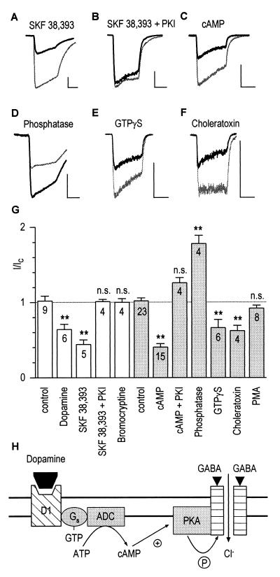
Figure 2.
Dopamine D1 receptors mediate down-modulation of GABAA receptors via phosphorylation by PKA in granule cells. (A) SKF-38393 (50 μM; see Fig. 1 for protocol) decreased the GABA control response (light trace) to 34% after 14 min of recording (dark trace). Horizontal and vertical scales in this figure correspond to 5 s and 500 pA, respectively. (B) The effect of SKF-38393 was abolished when PKA-inhibitory peptide (5 μg/ml) was included in the pipette. (C) Intracellular cAMP (100 μM) also decreased GABA-induced currents (I/IC = 0.65). (D) Potentiation of the GABA response to 186% of control by intracellular phosphatase (224 units/ml). (E–F) Decrease of currents by 100 μM GTP[γS] (I/IC = 0.56) and 1 μM choleratoxin (I/IC = 0.55). (G) Modulatory effects of extra- and intracellular drugs (open and shaded columns, respectively). The dashed line indicates the no-effect level. Statistical differences from control experiments are represented by asterisks (∗∗, P ≤ 0.01, n cells). (H) Model illustrating the sequence of events for the decrease of GABA-induced currents: Binding of dopamine to D1 receptors leads to activation of adenylate cyclase (ADC), production of cAMP, and eventually phosphorylation of GABAA receptors by PKA.

