
Figure 4. HSP90 inhibitors suppress B[a]P- and TCDD-mediated induction of CYP1A1 and CYP1B1 protein.
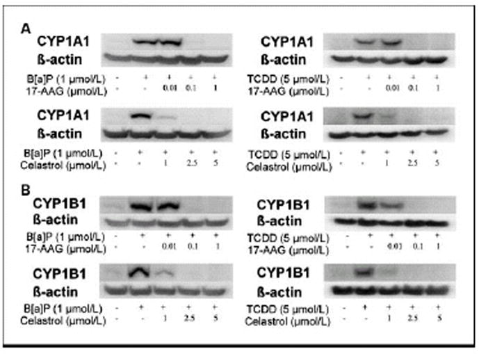
MSK-Leuk1 cells were treated with the indicated concentrations of 17-AAG or celastrol for 2 h. Subsequently, cells received vehicle, 1 μmol/L B[a]P or 5 nmol/L TCDD for 5 h, and were then harvested for Western blot analysis. Cellular lysate protein (100 μg/lane) was loaded onto a 10% SDS–polyacrylamide gel, electrophoresed and subsequently transferred onto nitrocellulose. Immunoblots were probed with antibodies specific for CYP1A1 (panel A), CYP1B1 (panel B) and β-actin.